 |
|
| Veterinary Article |
| An Unusual Reproductive Disorder in a Female Carpet Chameleon, Furcifer lateralis |
|
by Marc H. Kramer, DVM
|
|
This is a 3 year old female carpet chameleon (Furcifer lateralis) that had a long history of infertility and was found dead one morning on the cage bottom. This chameleon regularly laid clutches of infertile eggs over her lifetime despite being mated to a male on several occasions.
Over an 8 month period prior to her death, she had not laid any eggs as she typically would. A gradual increase in her abdominal size was noted over this time period, she appeared to have some difficulty maneuvering around her cage, and she was often seen resting her abdomen on branches in her enclosure. She had a good appetite and reasonably good attitude up until her death. No treatment or diagnostics were pursued while she was alive.
A necropsy was performed.
|
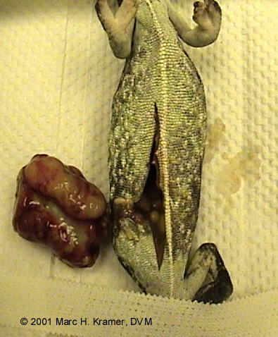
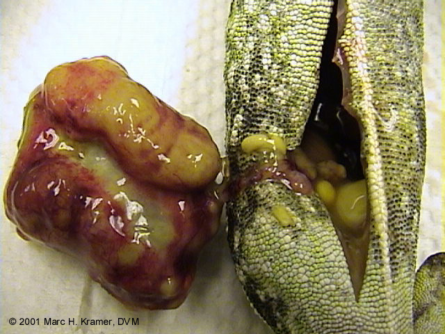
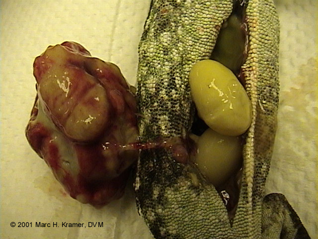
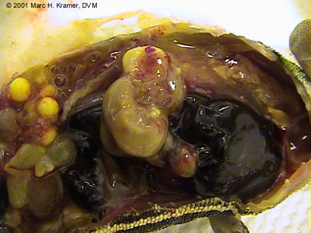
| The main finding on necropsy was a grossly distended and impacted oviduct (uterus), full of mummified eggs and inspissated yolk material. (Inspissated is defined as "being thickened, dried, or made less fluid by evaporation") Figures 1, 2, and 3 show the abnormal right oviduct which is severely enlarged and distorted due to the presence of old mummified dehydrated eggs. Mummified eggs were also found free-floating in the abdomen (coelom), which is also very abnormal as eggs should not normally be found outside of the oviduct. Follicles on the ovary may have had difficulty ovulating into the oviduct due to the multiple mummified eggs within it, and instead ovulated into the coelomic cavity (note: the coelom is the internal body cavity in reptiles and since reptiles do not have a true thorax and abdomen, there is only one continuous body cavity called the coelom.) |
 |
 |
 |
| Figure 1 |
Figure 2 |
Figure 3 |
|
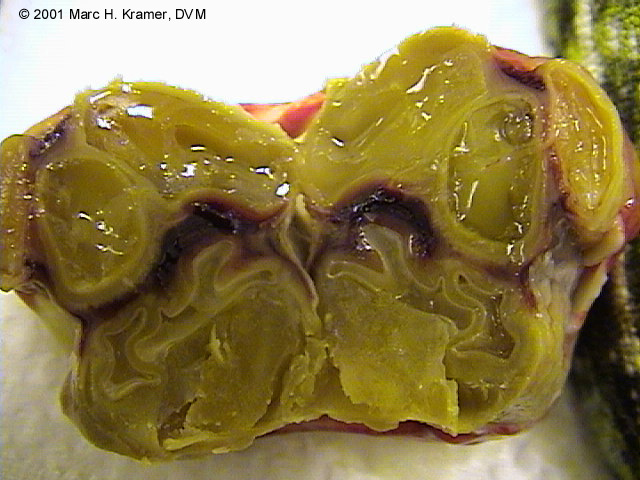
| Figure 4 shows a cut section of the abnormal oviduct, and it becomes clearly visible that the oviductal wall is massively distended, and the oviduct contains multiple mummified eggs and inspissated yolk. |
 |
| Figure 5 reveals several thoracic and abdominal structures, as well as two additional mummified eggs. In this picture, the head of the animal is to the right and the tail to the left. Several normal follicles are seen on the ovary (upper left). |
 |
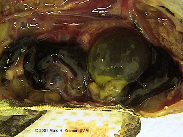
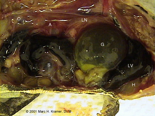
Figures 6 and 7 are the same image, but Figure 7 is labeled. A massively distended fluid-filled gallbladder seen on these images was another abnormal finding in this case and can be seen caudal (behind, or to the left in this picture) to the liver.
|
 |
 |
| Figure 6 |
Figure 7 |
|
|
KEY TO FIGURE 7:
|
| H = heart |
LN = lung |
S = spleen |
| LV = liver |
A = aorta |
O = ovary |
| G = gall bladder |
I = intestine |
|
|
| It is unknown why this animal originally developed this reproductive tract obstruction, or why she was infertile throughout her life. It is also unclear why the gall bladder was so massively distended. Histopathology (microscopic analysis of the tissues) and bacterial cultures may have helped to determine the cause but was not performed due to financial reasons. (the tissues have been preserved in formalin in the event that some generous person wants to donate the funds to pursue histopathology).
In retrospect, this chameleon should have been examined when the abdomen was observed to be enlarged and when egg production ceased. A radiograph (x-ray), ultrasound, or endoscopic examination may have helped to visualize the abnormal oviduct, and an ovariohysterectomy or hysterectomy could have been performed (albeit extremely challenging in a chameleon of this small size).
|
|
|
|
This page last updated on: Wednesday, November 27, 2002.
|
© 2002-2005 ADCHAM.com
ADCHAM logo illustrated by Randy Douglas. Web site design by Look Design, Inc.
Do not reproduce or redistibute any of content of this web site without express written permission from the authors.
|
|
|