 |
|
| Veterinary Article |
| Pilot Study on Endoscopic Techniques in Chameleons |
|
by Marc H. Kramer, DVM
|
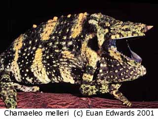 |
One chameleon species where endoscopic sex identification may be useful is the Meller's chameleon, Chamaeleo melleri (Figure 1). This species has been challenging to visually sex with accuracy. While the females tend to be slightly smaller than males, this is subtle and does not provide conclusive gender identification. Additionally, the medial crest anterior to the eyes is smaller in females and the dorsal crest and occipital lobes are less well developed; however, this is a distinction made only by experts. Recently, there has been discussion on sexing C. melleri by the presence or absence of a black spot on the back of the occipital flap (present in males, absent in females), yet this has not been a proven method and is not 100% reliable.
|
|
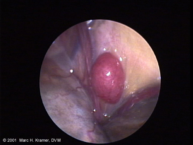
| Recently I have been employing endoscopic techniques for directly visualizing the gonads of C. melleri (Figure 2). This is turning out to be an important diagnostic tool not only in sexing Meller's chameleons, but also for sexing several species of monitor lizards, tortoises, and many other reptiles. |
|
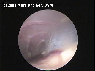
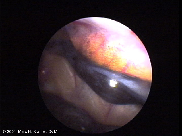
Figure 2. Testis (black) and epidydimis (white linear structure) of a C. melleri.
Fat (yellow mass) lies above the testis.
|
 |
|
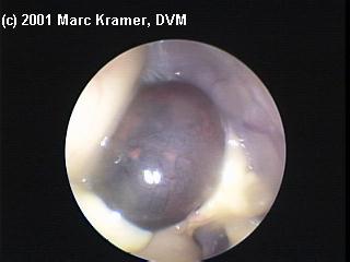
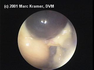
Brief anesthesia is needed for the procedure, typically a combination of injectable propofol and inhalant isoflurane gas. Once the animal is anesthetized, a small incision is made between the last two ribs, and the endoscope is placed inside the pleuroperitoneal cavity. From this point, the internal examination is performed, the gonad is visualized, and the animal can be accuately sexed. The entire procedure usually only takes 5-10 minutes and reveals an overwhelming amount of information including gender, health of various organs (Figures 3-6), and reproductive status (Figures 7-10)!
|
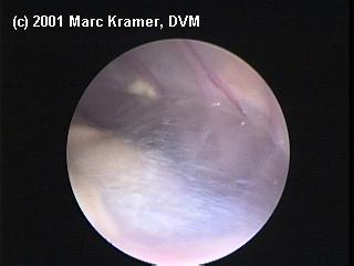
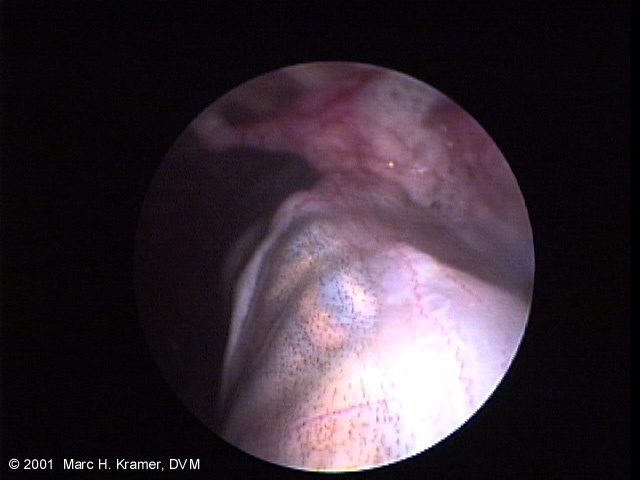
Figure 3. Stomach of C.melleri.
Close examination of this picture reveals a cricket inside the stomach! |
 |
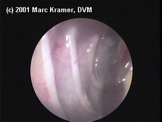
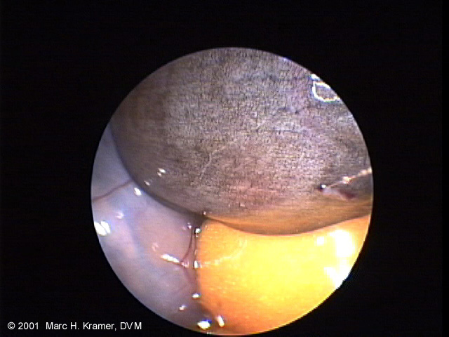
| Figure 4. Intestine (black) and fat (yellow). |
 |
| Figure 5. Liver and gastrointestinal tract. |
 |
| Figure 6. Spleen |
 |
|
|
|
|
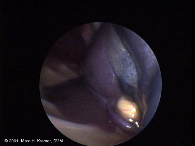
Figures 10 and 11. Abnormal egg (dark) amongst other egg material (white) within the coelom of a female C. calyptratus
|
 |
 |
|
|
|
|
|
|
|
This page last updated on: Wednesday, November 27, 2002
|
© 2002-2005 ADCHAM.com
ADCHAM logo illustrated by Randy Douglas. Web site design by Look Design, Inc.
Do not reproduce or redistibute any of content of this web site without express written permission from the authors.
|
|
|