 |
|
| Veterinary Article |
Metabolic Bone Disease with Emphasis on Pathologic
Limb Fractures in Chameleons |
|
by Marc H. Kramer, DVM
|
INTRODUCTION
Fractures of the limbs in chameleons are common, especially in young, rapidly growing animals. When this occurs, the front and/or hind limbs may appear bowed or have what appears to be an extra bend or joint in the leg. These fractures are typically Òpathologic fractures,Ó meaning they occur secondary to some disease process, rather than by acute trauma. In most cases, these pathologic fractures are a manifestation of a common reptile disorder called Metabolic Bone Disease (MBD).
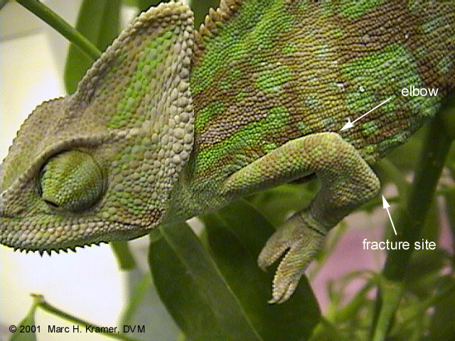
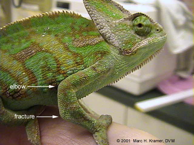
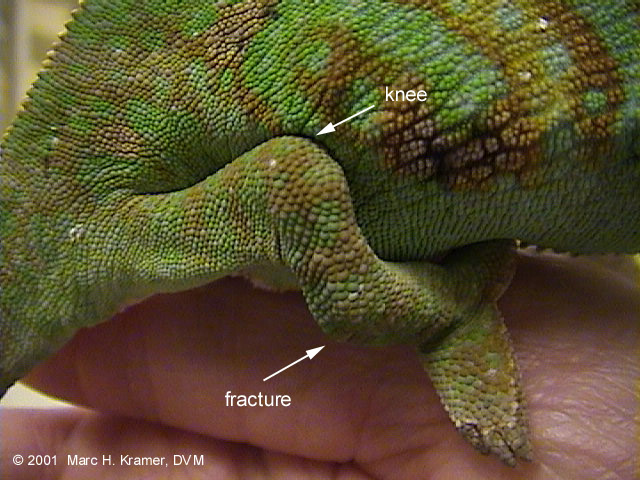
MBD is a complex disease process and is generally the long-term result of dietary deficiency of calcium or vitamin D, an unfavorable dietary calcium to phosphorus (Ca:P) ratio, and/or lack of exposure to ultraviolet B (UVB) light. The end result is weakened bone that easily breaks or bends. In chameleons, common fracture sites are in the front limbs midway between the elbow and the foot, and in the hind limbs midway between the knee and the foot. These areas are under a lot of weight-bearing tension and are very susceptible to MBD fractures. Other symptoms of MBD in chameleons include a soft, pliable, rubbery jaw, a bendable casque, and tongue dysfunction. In advanced cases, a chameleon may show inability to get up on its legs and lift its body off the ground (called lack of "truncal lifting"). The legs may move vigorously, yet the lizard is incapable of rising and ambulating. Eventually, there is a gradual decline in appetite, weight loss, lethargy, paralysis, seizures, and finally death if left untreated.
CASE REPORT:
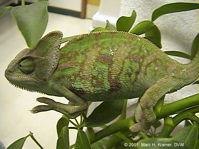
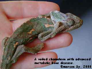
The juvenile veiled chameleons in the images to follow were from a clutch of about 35 affected animals. The entire clutch developed limb deformities as a result of a type of metabolic bone disease called rickets. Rickets is a disease of young growing animals caused by nutritional deficiencies, resulting in failure of normal calcification of bone. These animals were fed a large percentage of mealworms in the diet, which have a very poor Ca:P ratio. Additionally, the crickets that were being used were not regularly dusted with a calcium supplement. UVB lighting (ZooMed's ReptiSun 5.0) was in fact being used in this case but the animals still developed problems due to poor diet and inadequate calcium supplementation.
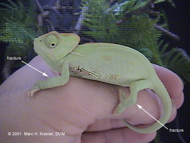
In the images below, observe the locations of the limb fractures which are the typical sites of MBD. These animals were actually in the recovery stage of the disease and the bones were beginning to solidify. Animals in this group had fractures affecting anywhere from 1 to 4 limbs:
|
|
|
|
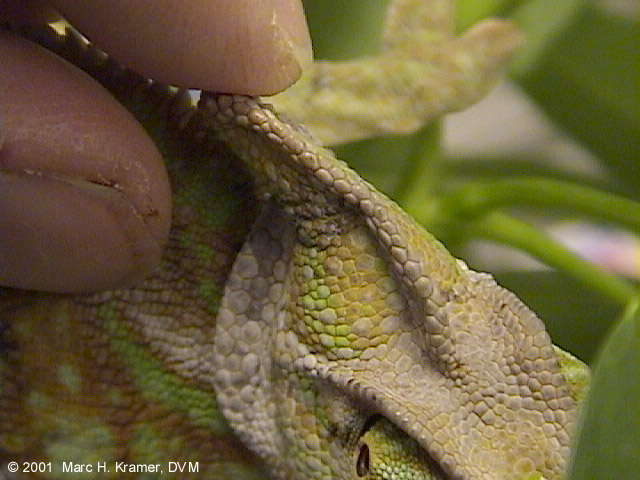
Note the very bendable rubbery casque in this animal:
|
|
|
|
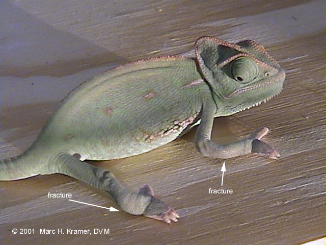
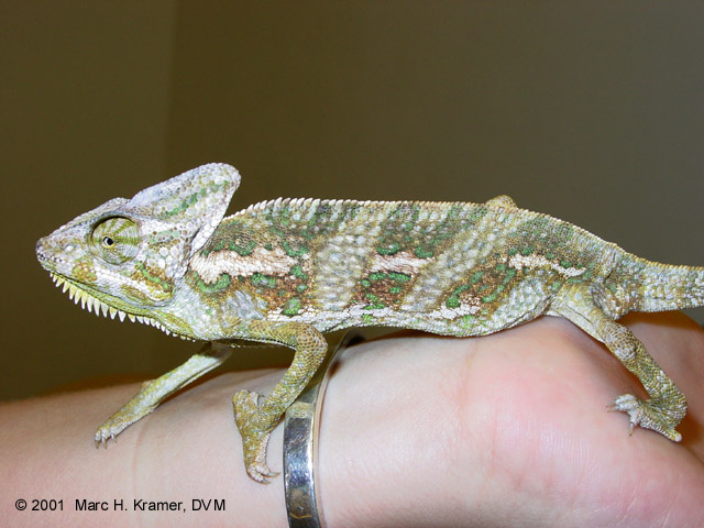
The animals depicted below were in the early to mid stages of the disease.
Notice both the fracture sites in the limbs, and the lack of truncal lifting.
|
|
|
| ADDITIONAL CASES
The following images were contributed by ADCHAM list members and reveal additional cases and effects of MBD in chameleons:
Severely stunted and emaciated 1 1/2 year old male veiled chameleon.
This animal weighed only 20 grams and was near death.
|
|
|
|
|
Advanced metabolic bone disease in a male veiled chameleon.
|
|
|
|
|
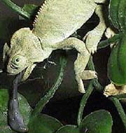
Limb abnormalities and tongue dysfunction in a chameleon (unidentified species).
|
|
|
|
|
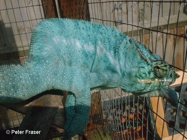
Spinal deformity, possibly MBD-related, in a male panther chameleon.
|
|
|
|
|
TREATMENT AND DIAGNOSIS
Treatment for MBD in chameleons must address several issues. The first is to correct proper nutrition. A varied diet of gut-loaded insects that are dusted regularly with calcium, especially for hatchlings and juveniles, is extremely important. Avoid biasing the diet with insects with a poor Ca:P ratio. Additionally, add appropriate UV lighting, and correct husbandry practices.
Treatment (under a veterinarian's supervision) generally involves administering medications which may include oral or injectable calcium, injectable vitamin D3, and/or calcitonin. Symptoms and specific medical problems will need to be addressed on a case-by-case basis, and may include procedures such as tube-feeding, correction of dehydration, or fracture stabilization. Be very careful handling patients with MBD as their bones are very fragile and subject to further injury if mishandled. Radiographs (X-rays) are useful to assess bone density and confirm suspicion of MBD, and measuring blood calcium and phosphorus levels can also aid diagnosis and help guide therapy. Those chameleons that recover from MBD and regain strong solid bones will probably still retain some hint of bumps or bends in the legs, which represent healed fractures. These animals can however go on to lead good quality lives. Sometimes other skeletal abnormalities may persist including general stunting, shortening of the mandible or maxilla, and spinal deviations. In general, MBD is a treatable disease if diagnosed and treated early.
|
|
|
|
This page last updated on: Wednesday, November 27, 2002
|
© 2002-2005 ADCHAM.com
ADCHAM logo illustrated by Randy Douglas. Web site design by Look Design, Inc.
Do not reproduce or redistibute any of content of this web site without express written permission from the authors.
|
|
|